What is Central Retinal Vascular Occlusion?
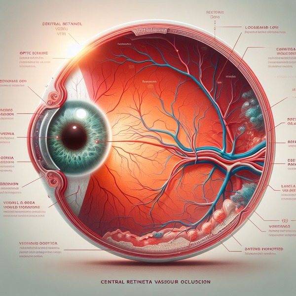
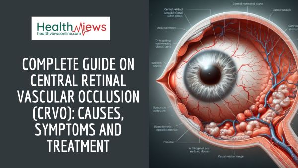
Central Retinal vascular occlusion (CRVO) is a partial or complete blockage of a vein draining blood from your retina. The retina is a layer of tissue in the back of the eye that aids in the translation of light into visible images. A retinal vein obstruction stops blood from leaving your retina. This can result in consequences such as increased ocular pressure and puffiness. Central Retinal vascular occlusion (CRVO) conditions require immediate attention to avoid or reduce vision loss.
There is currently no safe approach for clearing the vein. Treatment, on the other hand, can manage issues and protect your vision. Treatment is tailored to your specific needs by eye care specialists. To address your problem, you may require a combination of therapies ranging from injections to surgery. Source
Also, read Complete Guide on Age-Related Macular Degeneration: Causes, Symptoms, and Treatment

Causes
The most common cause of retinal vascular occlusion is artery hardening (atherosclerosis) and the production of a blood clot.
Blockage of smaller veins (branch veins or BRVO) in the retina happens frequently when thickened or hardened retinal arteries cross across and put pressure on a retinal vein.
Other eye disorders may result from retinal vascular blockage, such as:
- Glaucoma (high eye pressure) is caused by the formation of new, abnormal blood vessels at the front of the eye. Source
- Macular edema is caused by fluid leakage in the retina.
Symptoms
The symptoms of CRVO are easy to identify. They are as follows:
- Unexpected blindness in one of your eyes
- Sudden, total blurring of vision in one eye
- Over a few weeks, one eye gradually loses vision.
The effects could be felt for a few seconds or a few minutes. They could also be permanent. You most likely have branch retinal Vascular occlusion if you only experience partial blurring or loss of vision.
The symptoms of CRVO might resemble those of other medical conditions. Always seek the advice of your healthcare provider for a diagnosis.
Risk factors
Risk factors for retinal vascular occlusion include the following:
- Atherosclerosis
- Diabetes
- High blood pressure (hypertension)
Other eye diseases include glaucoma, macular edema, and vitreous haemorrhage.
As these conditions are more common in older adults, retinal vascular occlusion mostly affects these people.
Diagnosis
If your healthcare professional suspects you have CRVO, he or she will do an eye exam. First, your eye will be dilated. Various eye exams may be performed by your healthcare professional. These are performed to determine the type of blockage and the extent of damage. A fundoscopy is one eye test that can provide a definitive diagnosis. A biomicroscope and slit light are sometimes used for this procedure.
You will be tested for hypertension, glaucoma, and diabetes. If you are young, your doctor may want to see if your blood is thicker than usual. This can be accomplished by the use of a blood test known as a complete blood count, as well as other diagnostics. Other tests of your heart and blood vessel health may be performed by your healthcare professional to determine whether you have any other issues. These health issues could be related.
Treatment
There is no method to reverse or treat the blockage in your retinal vein right now. However, eye care specialists can avoid or treat the problems of retinal vein occlusion by using the following methods:
- Anti-VEGF injections – This is a protein that promotes the formation of new blood vessels (angiogenesis). A high level of VEGF can cause the creation of aberrant blood vessels, which can leak and cause edema. Source
- Steroid injections – Steroid medicine injections into your eye can also help minimize swelling. Steroid injections, on the other hand, can induce high blood pressure and cataracts in certain people. As a result, when anti-VEGF injections are ineffective, they are frequently used as a second-line treatment. Source
- Panretinal photocoagulation (PRP) – This laser procedure causes small burns in parts of your retina where blood flow is restricted. The quantity of proteins (VEGF) that encourage the creation of aberrant blood vessels is reduced as a result. Source
- Vitrectomy surgery – Posterior pars plana vitrectomy (PPV) is a surgery that helps persons with retinal vein occlusion who have: Source
- Severe eye bleeding (vitreous haemorrhage).
- Bleeding for more than four weeks.
- Bleeding that keeps returning.
- Retinal detachment.
- Medication to control risk factors – Many persons who have retinal vein occlusion have underlying illnesses such as hypertension, diabetes, or excessive cholesterol. These disorders can increase your chances of having blood vessel difficulties.
Prevention
Identifying and treating risk factors is the most effective strategy to prevent retinal vascular occlusion. Because retinal vascular occlusion is caused by vascular problems, it is critical to undertake lifestyle and nutritional adjustments to preserve your blood vessels and keep your heart healthy. These changes include:
- exercising
- losing weight or maintaining a healthy weight
- maintaining a nutritious diet free of saturated fat
- not smoking or quitting smoking
- controlling diabetes by maintaining a healthy blood sugar level
- taking aspirin or other blood thinners after discussing with your doctor
Routine doctor visits can help you determine whether you have any of the risk factors for retinal vascular occlusion. For example, if your doctor learns you have high blood pressure or diabetes, you can immediately begin preventive medication. Source
Also, read Complete Guide on Colour Blindness: Causes, Symptoms, and Diagnosis


