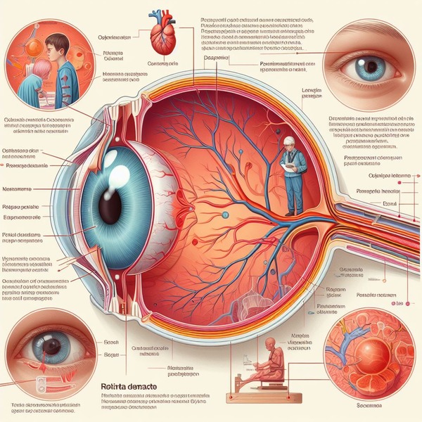
What is Retinal Detachment?
A painless but dangerous eye ailment is retinal detachment. It occurs when the retina, a layer of tissue in the back of your eye, separates from the tissues that support it. A detached retina impairs vision and can result in blindness.
Your retina detects light and delivers information to your brain, allowing you to see. Your retina loses blood flow when it moves away from the tissues that support it. These tissues’ blood arteries transport nutrients and oxygen to your retina. Sudden changes, such as eye floaters flashes and dimming side vision, are indicators that something is happening. A detached retina requires immediate treatment.
Also, Read Complete Guide on Age-Related Macular Degeneration: Causes, Symptoms, and Treatment
Causes
Retinal detachment occurs when the portion of the eye responsible for image creation peels away from the rear of the eye. It can be caused by an injury, inflammation, damage, or structural changes to the eye over time. When an image concentrates on the retina, nerve cells process the information and transfer it to the brain via electrical impulses.
Damage to the retina can impair a person’s vision. When this layer slips away from its normal place, retinal detachment occurs. Small tears in the retina can occasionally cause detachment. The macula is the region of the retina that controls vision. The macula may or may not become detached during retinal detachment. If it does, the risk of central vision loss increases.

Risk factors
Anyone can have retinal detachment, but there are a few factors that may increase your risk. These include:
- family history of retinal detachment
- you’ve previously suffered a major eye injury
- you’ve had eye surgery in the past (to treat cataracts, for example)
- you’ve been diagnosed with certain eye conditions
- you’re extremely nearsighted
- ageing
Visual disease and other visual disorders may increase your chance of retinal detachment. These eye problems may include:
- Diabetic retinopathy (a condition in which blood vessels in the retina are affected by diabetes)
- Posterior vitreous Detachment (the gel-like fluid in the centre of the eye pushes away from the retina)
- Retinoschisis (division of the retina into two layers)
- Lattice degeneration (retinal thinning)
Symptoms
The detachment of the retina is not painful. However, warning signals nearly always arise before the event begins or has progressed, such as:
- The unexpected emergence of a large number of floaters – specks that appear to move across your field of vision
- One or both eyes flash with light (photopsia)
- Vision distortion
- Reduced peripheral (side) vision gradually
- A curtain-like shadow across your field of vision.
Diagnosis
An eye exam is required to determine retinal detachment. A dilated eye exam will be performed by your eye care physician to examine your retina. After the dilated eye exam, your provider may request additional tests. These are non-invasive tests. They will not cause any harm. They allow your provider to see your retina more clearly and in greater detail:
- Optical coherence tomography (OCT): This imaging usually requires dilating eye drops. Then you position yourself in front of the OCT machine. The equipment scans your eye without touching it.
- Fundus imaging: Your provider might take wide-angle images of your retina during fundus imaging. For this test, your provider would normally dilate your pupils.
- Eye (ocular) ultrasound: You will not require dilating drops for this scan, however, your practitioner may use drops to numb your eyes so you are not bothered. To scan your eye, your physician gently presses equipment against the front of it.
Treatment
To heal a retinal tear, hole, or detachment, surgery is nearly always required. There are several methods accessible.
Retinal tears
If a retinal tear or hole has not yet proceeded to detachment, your eye surgeon may recommend one of the following procedures to avoid detachment and maintain vision.
- Laser surgery (photocoagulation) – It involves the surgeon directing a laser beam through the pupil into the eye.
- Freezing (cryopexy) – After numbing your eye with a local anaesthetic, the surgeon places a freezing probe on the outer surface of the eye directly above the tear.
Retinal detachment
If your retina has detached, you’ll require surgery to restore it as soon as possible.
- Injecting air or gas into your eye – During a pneumatic retinopexy procedure, the surgeon inserts a gas or air bubble into the vitreous cavity, the central region of the eye.
- Indenting the surface of your eye – During a process known as scleral buckling, the surgeon will stitch, or suture, a piece of silicone material to the afflicted portion of your eye’s white, or sclera.
- Draining and replacing the fluid in the eye – During this vitrectomy surgery, the surgeon excises the vitreous and any tissue that is exerting pressure on the retina.
Diet
- Dark green leafy vegetables including Spinach, Kale Mustard Greens, Collard Greens, and Chard.
- Add Vitamin C rich fruits including oranges, sweet lime, grapes etc.
- Nuts like walnuts, almonds, hazelnuts etc.
Prevention
In most cases, there is no way to avoid retinal detachment. However, you can take precautions to avoid retinal detachment caused by injury or disease. These include:
- wear protective eyewear when participating in sports, performing heavy lifting, or utilising tools
- if you have diabetes, keep your blood sugar under control
- get dilated eye tests regularly
It’s also important to understand the symptoms of retinal detachment and to see your doctor as soon as you notice them.
Also, Read Complete Information about Cataracts: Causes, Symptoms and Treatment


